Beranda
/ Upper Back Anatomy / Human Anatomy Showing Deep Muscles In The N Stocktrek Images Icanvas / The superficial and intermediate muscles do not develop in the back, and are classified as extrinsic muscles.
Upper Back Anatomy / Human Anatomy Showing Deep Muscles In The N Stocktrek Images Icanvas / The superficial and intermediate muscles do not develop in the back, and are classified as extrinsic muscles.
Insurance Gas/Electricity Loans Mortgage Attorney Lawyer Donate Conference Call Degree Credit Treatment Software Classes Recovery Trading Rehab Hosting Transfer Cord Blood Claim compensation mesothelioma mesothelioma attorney Houston car accident lawyer moreno valley can you sue a doctor for wrong diagnosis doctorate in security top online doctoral programs in business educational leadership doctoral programs online car accident doctor atlanta car accident doctor atlanta accident attorney rancho Cucamonga truck accident attorney san Antonio ONLINE BUSINESS DEGREE PROGRAMS ACCREDITED online accredited psychology degree masters degree in human resources online public administration masters degree online bitcoin merchant account bitcoin merchant services compare car insurance auto insurance troy mi seo explanation digital marketing degree floridaseo company fitness showrooms stamfordct how to work more efficiently seowordpress tips meaning of seo what is an seo what does an seo do what seo stands for best seotips google seo advice seo steps, The secure cloud-based platform for smart service delivery. Safelink is used by legal, professional and financial services to protect sensitive information, accelerate business processes and increase productivity. Use Safelink to collaborate securely with clients, colleagues and external parties. Safelink has a menu of workspace types with advanced features for dispute resolution, running deals and customised client portal creation. All data is encrypted (at rest and in transit and you retain your own encryption keys. Our titan security framework ensures your data is secure and you even have the option to choose your own data location from Channel Islands, London (UK), Dublin (EU), Australia.
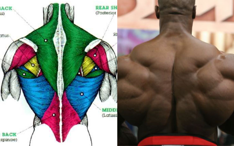
Upper Back Anatomy / Human Anatomy Showing Deep Muscles In The N Stocktrek Images Icanvas / The superficial and intermediate muscles do not develop in the back, and are classified as extrinsic muscles.. The upper back muscles of the rhomboids and the trapezius are responsible for many of the movements of the scapula which in turn plays a huge role in the stability and mobility of the shoulder. It originates from the base of the skull, along the nuchal ligament and the 7th cervical vertebra , which is that bony landmark on the back of your neck. There is a set of muscles in the upper back (called the thoracic area) called the spinalis thoracis. This is my video about the muscles of the back. The trapezius and latissimus dorsi muscles connect the upper limb to the vertebral column.
It runs from the neck to the upper back. Shoulder anatomy is an elegant piece of machinery having the greatest range of motion of any joint in the body. Upper back pain is most commonly caused by muscle irritation or tension, also called myofascial pain. The main superficial muscles of the back are the following: The cervical spine supports the weight and movement of your head and protects the nerves exiting your brain.
How To Draw The Upper Back Anatomy And Motion Proko from thumbs.gfycat.com These include the cervical vertebrae in the neck, the thoracic vertebrae of the ribcage in the upper and middle back, the lumbar vertebrae in the lower back, and the vertebrae that are part of the pelvis. The muscles of the chest and upper back occupy the thoracic region of the body inferior to the neck and superior to the abdominal region and include the muscles of the shoulders. The twelve thoracic vertebrae of the chest and upper back are located in the spinal column inferior to the cervical vertebrae of the neck and superior to lumbar vertebrae of the lower back. The complexity of this region means that dysfunction can occur either due to injury or progressive pain and degeneration. License image the deltoid, teres major, teres minor, infraspinatus, supraspinatus (not shown) and subscapularis muscles (not shown) all extend from the scapula to the humerus and act on the shoulder joint. Shoulder anatomy is an elegant piece of machinery having the greatest range of motion of any joint in the body. The traps) the latissimus dorsi (a.k.a. The nervous system of the thorax is a vital part of the nervous system as a whole, as it includes the spinal cord, peripheral nerves, and autonomic ganglia that communicate with and control many vital organs.
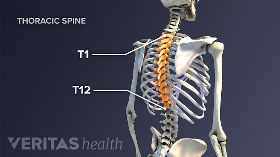
It is like that for several reasons, all of which you can understand by looking at the anatomy of the thoracic spine.
It comprises the vertebral column (spine) and two compartments of back muscles; These include the cervical vertebrae in the neck, the thoracic vertebrae of the ribcage in the upper and middle back, the lumbar vertebrae in the lower back, and the vertebrae that are part of the pelvis. The iliocostalis muscles are furthest from the spine. The cause may be poor posture (such as forward head posture) or any type of irritation of the large back and shoulder muscles, including muscle strain or spasms. See upper back stock video clips. This muscle is located on the upper portion of the back anatomy, underneath the trapezius. The traps) the latissimus dorsi (a.k.a. Upper back pain is most commonly caused by muscle irritation or tension, also called myofascial pain. Upper back pain rear view of spine back pain spine sports spine spine surgery spine white background back ache x human anatomy illustration human anatomy on white background upper body stretch. The superficial and intermediate muscles do not develop in the back, and are classified as extrinsic muscles. It runs from the neck to the upper back. The rhomboid muscle is activated as you bring and squeeze your scapula or shoulder blades back and together. The complexity of this region means that dysfunction can occur either due to injury or progressive pain and degeneration.
As viewed from the side, the cervical spine forms a lordotic curve by gently curving toward the front of the body and then back. These include the cervical vertebrae in the neck, the thoracic vertebrae of the ribcage in the upper and middle back, the lumbar vertebrae in the lower back, and the vertebrae that are part of the pelvis. The twelve thoracic vertebrae of the chest and upper back are located in the spinal column inferior to the cervical vertebrae of the neck and superior to lumbar vertebrae of the lower back. Some of the exercises which an individual can do to relax or loosen the upper neck and back muscles are: The iliocostalis muscles are furthest from the spine.
Grow Your Upper Back With This Workout Spotmebro Com from spotmebro.com The rhomboid muscle is activated as you bring and squeeze your scapula or shoulder blades back and together. Before giving our recommendations for upper back exercises, it's important to first go over the anatomy of the back musculature. The complexity of this region means that dysfunction can occur either due to injury or progressive pain and degeneration. The back functions are many, such as to house and protect the spinal cord, hold the body and head upright, and adjust the movements of the upper and lower limbs. Some of the exercises which an individual can do to relax or loosen the upper neck and back muscles are: The cervical spine is the top part of the spine. It runs from the neck to the upper back. The cervical spine protects the nerves connecting to.
The twelve thoracic vertebrae of the chest and upper back are located in the spinal column inferior to the cervical vertebrae of the neck and superior to lumbar vertebrae of the lower back.
There is a set of muscles in the upper back (called the thoracic area) called the spinalis thoracis. Chart human nervous system 13 photos of the chart human nervous system human body nervous system, human nervous system definition, human nervous system diagram, human nervous system diseases, human nervous system facts, human nervous system for kids, human nervous system function, human nervous system pdf, human. It runs from the neck to the upper back. The deep muscles develop embryologically in the back, and are thus described as intrinsic muscles. It originates from the base of the skull, along the nuchal ligament and the 7th cervical vertebra , which is that bony landmark on the back of your neck. Related posts of upper back muscle diagram muscle anatomy app. Upper back pain rear view of spine back pain spine sports spine spine surgery spine white background back ache x human anatomy illustration human anatomy on white background upper body stretch. The cause may be poor posture (such as forward head posture) or any type of irritation of the large back and shoulder muscles, including muscle strain or spasms. As viewed from the side, the cervical spine forms a lordotic curve by gently curving toward the front of the body and then back. The rhomboid muscle is activated as you bring and squeeze your scapula or shoulder blades back and together. These include the cervical vertebrae in the neck, the thoracic vertebrae of the ribcage in the upper and middle back, the lumbar vertebrae in the lower back, and the vertebrae that are part of the pelvis. A band of pain just under the shoulder blades is also common. Injuries to these muscles can cause upper back pain.
Related posts of upper back muscle diagram muscle anatomy app. Upper right back pain under shoulder blade. A band of pain just under the shoulder blades is also common. Upper back pain rear view of spine back pain spine sports spine spine surgery spine white background back ache x human anatomy illustration human anatomy on white background upper body stretch. Try the injurymap exercise app now.
All About Upper Back Pain from embed.widencdn.net It is very stiff, and the thoracic spine has a limited range of motion. The upper back originates at the base of your neck, incorporates both shoulders and extends down to mid spine, including your ribs. It originates from the base of the skull, along the nuchal ligament and the 7th cervical vertebra , which is that bony landmark on the back of your neck. Back anatomy the back is the body region between the neck and the gluteal regions. The back functions are many, such as to house and protect the spinal cord, hold the body and head upright, and adjust the movements of the upper and lower limbs. The upper back muscles of the rhomboids and the trapezius are responsible for many of the movements of the scapula which in turn plays a huge role in the stability and mobility of the shoulder. Upper back pain is most commonly caused by muscle irritation or tension, also called myofascial pain. The trapezius and latissimus dorsi muscles connect the upper limb to the vertebral column.
Thoracic vertebrae interlock tightly by overlapping their spinous processes, giving stability to the spine in this region.
The top of the cervical spine connects to the skull, and the bottom connects to the upper back at about shoulder level. The traps) the latissimus dorsi (a.k.a. The upper portion is that trapezoid shape visible from the front of the body. The muscles of the chest and upper back occupy the thoracic region of the body inferior to the neck and superior to the abdominal region and include the muscles of the shoulders. It comprises the vertebral column (spine) and two compartments of back muscles; Looking for a solution to your back pain problem? Muscle strain, sprains, and spasms can affect the rhomboid muscles, which are located in the middle of the shoulder blades.this pain is mostly felt in. The main superficial muscles of the back are the following: Upper back pain rear view of spine back pain spine sports spine spine surgery spine white background back ache x human anatomy illustration human anatomy on white background upper body stretch. The back functions are many, such as to house and protect the spinal cord, hold the body and head upright, and adjust the movements of the upper and lower limbs. The upper back originates at the base of your neck, incorporates both shoulders and extends down to mid spine, including your ribs. It originates from the base of the skull, along the nuchal ligament and the 7th cervical vertebra , which is that bony landmark on the back of your neck. The nervous system of the thorax is a vital part of the nervous system as a whole, as it includes the spinal cord, peripheral nerves, and autonomic ganglia that communicate with and control many vital organs.